neuroanatomy through clinical cases pdf
Neuroanatomy Through Clinical Cases: A Comprehensive Overview
Hal Blumenfeld’s resource, alongside texts like Paul Young’s Neuroanatomia Clinica, offers a case-based approach to understanding complex neurological structures.
PDF versions, such as blumenfeld_h_neuroanatomy_through_clinical_case.pdf (72.5 MB, 2015), are widely available, facilitating focused study of clinical correlations.
Hal Blumenfeld’s Neuroanatomy Through Clinical Cases stands as a pivotal text for medical students, residents, and practitioners seeking a deeper comprehension of the nervous system. Unlike traditional, purely anatomical approaches, this resource uniquely integrates clinical scenarios with neuroanatomical principles, fostering a more intuitive and lasting understanding.
The availability of the text in PDF format – exemplified by files like blumenfeld_h_neuroanatomy_through_clinical_case.pdf (a 72.5 MB file last updated October 14, 2015) – significantly enhances accessibility for learners. This digital format allows for convenient study, annotation, and portability.
Furthermore, the book’s emphasis on clinical correlation, as seen in related works like Gould D.J.’s Clinical Neuroanatomy: A Case-Based Approach, bridges the gap between theoretical knowledge and real-world neurological presentations. It’s a valuable tool for diagnosing and localizing lesions, understanding neurological disorders, and interpreting neuroimaging findings.
Hal Blumenfeld’s “Neuroanatomy Through Clinical Cases”
Hal Blumenfeld’s work is distinguished by its deliberate focus on applying neuroanatomical knowledge to actual patient presentations. The second edition builds upon the strengths of the first, offering a robust collection of clinical cases designed to challenge and reinforce understanding of neurological structures and pathways.
The readily available PDF version (blumenfeld_h_neuroanatomy_through_clinical_case.pdf, 72.5 MB) facilitates detailed study, allowing users to navigate the text efficiently and revisit complex concepts. This digital accessibility is crucial for modern medical education.
The book’s methodology encourages a diagnostic approach, prompting readers to localize lesions based on clinical findings – a skill essential for neurologists and neurosurgeons. It complements other resources like Gerald’s Clinical Neuroanatomy and Neuroscience, providing a practical, case-driven perspective on neuroanatomy.
The Importance of Clinical Correlation in Neuroanatomy
Traditional neuroanatomy instruction often focuses on rote memorization of structures, but true understanding requires linking anatomy to clinical presentation. Resources like Hal Blumenfeld’s “Neuroanatomy Through Clinical Cases” – available as a PDF – exemplify this crucial connection.
Understanding how lesions in specific brain regions manifest as distinct neurological deficits is paramount. Clinical cases force students to actively apply their anatomical knowledge, enhancing retention and diagnostic skills. This approach moves beyond theoretical knowledge to practical application.
Furthermore, correlating neuroanatomy with clinical findings aids in accurate localization, essential for differential diagnosis. Texts like Taylor’s and Gould’s emphasize this case-based learning, mirroring the real-world challenges faced by clinicians. Accurate diagnosis, even in complex cases like autistic regression with epileptiform activity, relies on this foundation.

Core Concepts & Anatomical Regions
PDF resources, including Blumenfeld’s work, detail brainstem, cerebellar, and basal ganglia functions, linking anatomical structures to observed clinical dysfunctions and syndromes.
Brainstem Lesions and Clinical Presentation
Clinical cases, often detailed in PDF formats like Hal Blumenfeld’s text, illustrate how lesions within the brainstem manifest neurologically.
Damage to specific brainstem nuclei impacts cranial nerve function, leading to deficits in areas like eye movement, facial sensation, or swallowing.
Neuroanatomy through clinical cases emphasizes correlating lesion location with observed symptoms; for example, a lesion affecting the midbrain can cause pupillary abnormalities.
Understanding the decussation of fibers within the brainstem is crucial, as lesions often produce contralateral symptoms below the level of the lesion.
Resources highlight syndromes like Wallenberg’s syndrome (lateral medullary syndrome) resulting from vertebral artery occlusion, causing ipsilateral ataxia and contralateral pain/temperature loss.
Detailed PDF analyses demonstrate how precise anatomical knowledge is essential for accurate diagnosis and localization of brainstem pathology, guiding appropriate clinical management.
Cerebellar Dysfunction: Case Studies
Neuroanatomy through clinical cases, readily available in PDF resources like Blumenfeld’s work, showcases cerebellar dysfunction through illustrative patient presentations.
Case studies demonstrate how lesions affecting the cerebellum – including the vermis, hemispheres, and peduncles – result in characteristic impairments.
These impairments commonly include ataxia (loss of coordination), dysmetria (inaccurate movements), and intention tremor, observable during finger-to-nose testing.
PDF analyses often detail how specific cerebellar regions contribute to distinct motor functions; for instance, vermian lesions cause truncal ataxia.
Furthermore, cases highlight non-motor deficits, such as cognitive and emotional disturbances, linked to cerebellar involvement.
Understanding the cerebellar circuitry – its inputs from the spinal cord and cerebral cortex, and outputs to the thalamus – is vital for interpreting clinical findings presented in these detailed PDF case reports.
Basal Ganglia Disorders: A Clinical Approach
Neuroanatomy through clinical cases, often found in accessible PDF formats like Blumenfeld’s text, emphasizes a practical approach to basal ganglia disorders.
These resources present cases illustrating the clinical consequences of dysfunction within the caudate, putamen, globus pallidus, substantia nigra, and subthalamic nucleus.
PDF analyses detail how lesions manifest as movement disorders – Parkinsonism (rigidity, tremor, bradykinesia), Huntington’s disease (chorea), and dystonia.
Clinical correlation is key; understanding the direct and indirect pathways within the basal ganglia helps localize the lesion based on observed symptoms.
For example, PDF case studies demonstrate how subthalamic nucleus lesions cause hemiballismus, a violent flinging movement.
Furthermore, these resources explore the cognitive and behavioral aspects of basal ganglia dysfunction, highlighting the interconnectedness of neural circuits, all detailed within the PDF.

Specific Clinical Cases & Neurological Disorders
Neuroanatomy through clinical cases, often available as a PDF, illustrates stroke syndromes, epilepsy, and multiple sclerosis, linking anatomy to observed clinical manifestations.
Stroke Syndromes: Localizing Lesions

Neuroanatomy Through Clinical Cases, frequently accessed as a PDF resource, excels at demonstrating how specific stroke syndromes correlate with precise lesion locations within the brain.
Understanding vascular territories and their corresponding neurological deficits is paramount; the text utilizes case studies to illustrate this principle. For instance, lesions affecting the middle cerebral artery often present with contralateral hemiparesis and sensory loss, alongside aphasia if the dominant hemisphere is involved.
Conversely, anterior cerebral artery strokes typically cause leg weakness disproportionately. Posterior cerebral artery involvement frequently manifests as visual field defects.
The PDF format allows for detailed review of neuroimaging alongside clinical presentations, reinforcing the crucial link between anatomical knowledge and accurate lesion localization. This approach is vital for effective diagnosis and treatment planning, enabling clinicians to predict potential deficits based on the affected brain region.
Furthermore, the resource emphasizes the importance of considering variations in arterial anatomy, as collateral circulation can influence the clinical picture.
Epilepsy and Neuroanatomical Substrates
Neuroanatomy Through Clinical Cases, often consulted in PDF format, highlights the critical relationship between epileptiform activity and underlying neuroanatomical structures. The resource details how seizure semiology – the observed clinical manifestations – directly reflects the origin and spread of abnormal electrical discharges.
Temporal lobe epilepsy, for example, frequently arises from the hippocampus and amygdala, manifesting as complex partial seizures with automatisms and altered awareness. Frontal lobe seizures can present with bizarre motor phenomena due to involvement of motor and premotor cortices.
The PDF allows for detailed examination of neuroimaging findings, correlating structural abnormalities (like hippocampal sclerosis) with seizure patterns.
Furthermore, the text addresses autistic regression linked to epileptiform activity, emphasizing the importance of EEG-video monitoring for accurate diagnosis. Understanding these neuroanatomical substrates is crucial for targeted treatment strategies, including surgical resection or neuromodulation.
Overdiagnosis of epilepsy (2025 of all new cases) underscores the need for precise localization.
Multiple Sclerosis: Demyelination and Clinical Manifestations
Neuroanatomy Through Clinical Cases, accessible as a PDF resource, elucidates the pathophysiology of Multiple Sclerosis (MS) by linking demyelination to specific clinical presentations. The text emphasizes that MS lesions, often visualized through neuroimaging, disrupt neuronal pathways throughout the central nervous system.
Optic neuritis, a common initial symptom, results from demyelination of the optic nerve, leading to vision loss. Lesions in the spinal cord can cause weakness, spasticity, and sensory disturbances. Brainstem involvement manifests as ataxia, diplopia, and nystagmus.
The PDF format facilitates detailed study of lesion patterns and their correlation with disability. Understanding the neuroanatomical distribution of MS lesions is vital for predicting disease progression and tailoring rehabilitation strategies.
Furthermore, the resource highlights the variability in clinical manifestations, reflecting the unpredictable nature of demyelination and axonal damage.

Diagnostic Tools & Techniques
Neuroimaging, including PDF-referenced techniques, is crucial; ultrasound assesses peripheral nerves, while neurophysiology aids lesion localization, as detailed in available resources.
Neuroimaging in Clinical Neuroanatomy
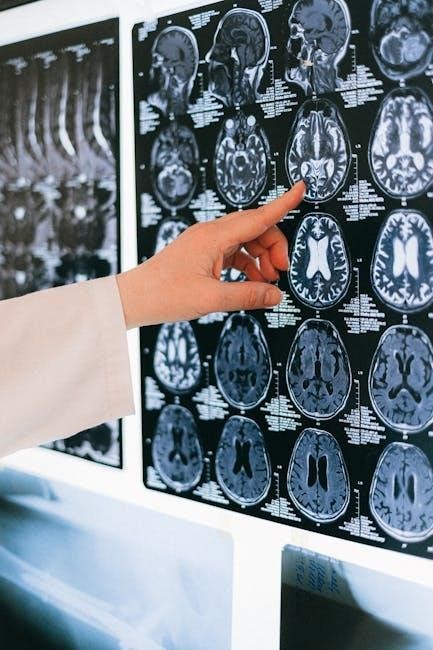
Neuroimaging plays a pivotal role in correlating anatomical structures with clinical presentations, a core tenet of resources like Hal Blumenfeld’s work and related PDF materials such as Neuroanatomia Clinica by Paul Young.
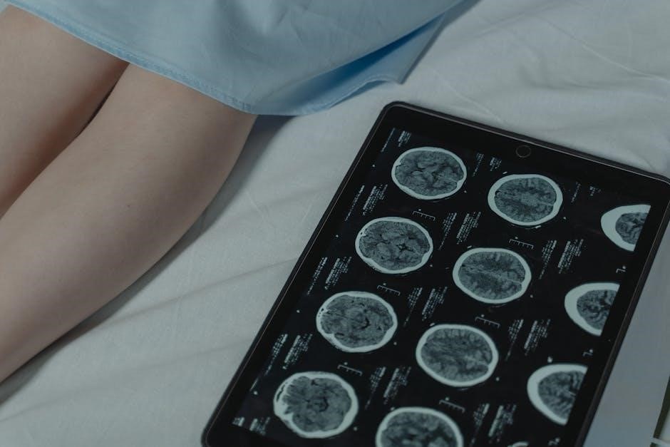
Techniques like MRI and CT scans allow visualization of lesions impacting the brainstem, cerebellum, and basal ganglia, aiding in the diagnosis of stroke syndromes, epilepsy, and multiple sclerosis.
The ability to pinpoint lesion location is essential, especially when considering conditions like autistic regression and associated epileptiform activity, as detailed in research exploring neuroanatomical substrates.
Furthermore, understanding neuroanatomy is vital for interpreting neuroimaging findings in cases of chronic pain and executive function disorders, enabling a more precise diagnostic approach.
Ultrasound in Peripheral Nerve Assessment
While resources like Hal Blumenfeld’s Neuroanatomy Through Clinical Cases and associated PDF documents primarily focus on central nervous system imaging, peripheral nerve assessment benefits significantly from techniques like ultrasound.
As highlighted in available data, ultrasound allows for dynamic visualization of nerves, aiding in the diagnosis of neuropathies and entrapment syndromes. This complements the static anatomical views provided by traditional neuroimaging.
Specifically, the obturator nerve can be effectively assessed using ultrasound, as demonstrated in documented cases. This is particularly useful when correlating anatomical variations with clinical presentations of pain or weakness.
Integrating ultrasound findings with a strong foundation in neuroanatomy – gained from resources like Paul Young’s work – enhances diagnostic accuracy and guides targeted interventions for peripheral nerve disorders.
The Role of Neurophysiology in Localization
Understanding neurophysiology is crucial when utilizing resources like Hal Blumenfeld’s Neuroanatomy Through Clinical Cases and related PDF materials for accurate lesion localization. While anatomical knowledge, derived from texts such as Paul Young’s Neuroanatomia Clinica, provides the ‘where’, neurophysiology explains the ‘how’.
Clinical data suggests a potential for overdiagnosis of epilepsy (around 20% of new cases), emphasizing the need to correlate clinical symptoms with objective neurophysiological findings. This involves techniques like EEG and evoked potentials.
By integrating neuroanatomy and neurophysiology, clinicians can refine diagnoses and tailor treatment strategies. This approach is particularly vital in complex cases involving functional disturbances or subtle anatomical lesions.
Ultimately, a combined understanding of structure and function, facilitated by resources and advanced diagnostic tools, optimizes patient care and improves neurological outcomes.

Advanced Topics & Future Directions
Neuroanatomy, coupled with clinical data from resources like the PDF versions of Blumenfeld’s work, informs research into chronic pain and executive function disorders.
Neuroanatomy and Chronic Pain
Neuroanatomical understanding is crucial when investigating the complexities of chronic pain, as highlighted within resources like Hal Blumenfeld’s Neuroanatomy Through Clinical Cases – often accessed as a PDF.
The literature suggests a mixed model for chronic pain development, involving neuropathic sensitive phenomena and enhanced central sensitization (CS). Clinical cases demonstrate how anatomical lesions, even subtle ones, can dramatically alter pain pathways.
Detailed PDF resources allow for focused study on specific nerve involvement, such as the obturator nerve, and how its dysfunction manifests clinically. Furthermore, understanding the neurophysiological basis of pain, alongside anatomical knowledge, is vital for effective treatment strategies.
Research explores how functional neuroanatomy contributes to the weight of chronic pain experiences, necessitating a comprehensive approach integrating anatomical and physiological insights derived from detailed case studies and readily available PDF materials.
Executive Function Disorders and Neuroanatomical Basis
Executive function deficits are intricately linked to specific brain regions, a connection thoroughly explored in resources like Hal Blumenfeld’s Neuroanatomy Through Clinical Cases, frequently consulted in PDF format.
Understanding the neuroanatomical substrates is essential for assessing and rehabilitating these disorders. Case studies detailed in available PDF materials demonstrate how damage to the prefrontal cortex, and its connections, impacts planning, decision-making, and working memory.
Clinical assessments, informed by neuroanatomical knowledge, help localize the site of dysfunction. Resources like Gould’s Clinical Neuroanatomy: A Case-Based Approach, complement Blumenfeld’s work, offering practical application of these principles.
The assessment and rehabilitation strategies are significantly enhanced by a firm grasp of the underlying neuroanatomy, readily accessible through detailed case presentations found within comprehensive PDF guides.
Autistic Regression and Epileptiform Activity
The connection between autistic regression and epileptiform activity is a complex area, often investigated using resources like Hal Blumenfeld’s Neuroanatomy Through Clinical Cases, available as a detailed PDF document.
In certain instances, particularly those involving clinical seizures, a diagnosis of autistic epileptiform regression is considered. Understanding the underlying neuroanatomy is crucial for differentiating these conditions and identifying potential seizure foci.
PDF versions of neuroanatomy texts aid in visualizing the brain regions involved, allowing for a more nuanced understanding of how epileptiform discharges might contribute to developmental regression.

Clinical cases, often presented in PDF format, illustrate the importance of correlating neurophysiological findings with anatomical structures, ultimately guiding diagnosis and treatment strategies for these challenging presentations.